Reimbursement guidelines for MESI mTABLET in the USA
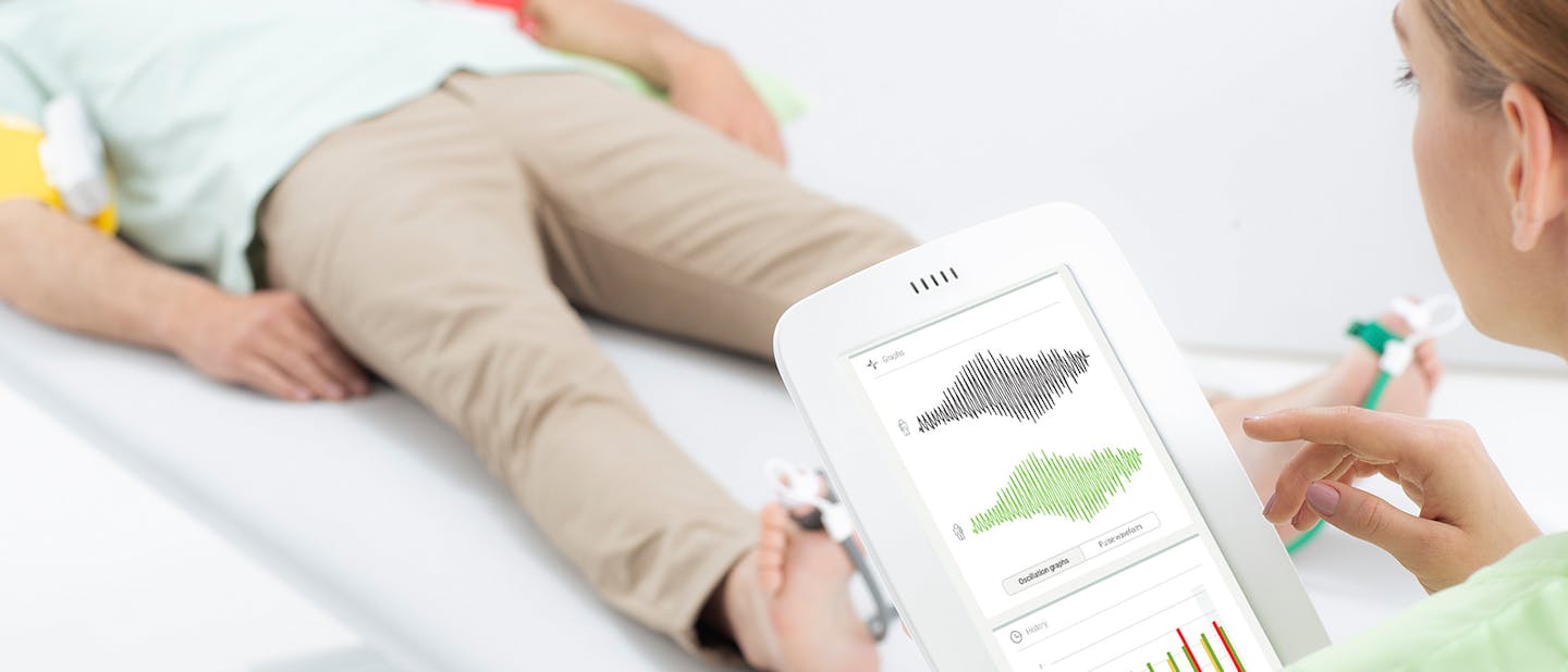
Automated ABI/TBI with MESI mTABLET: Reimbursable under CPT 93922
For clinicians: The automated MESI mTABLET system delivers guideline-concordant ABI/TBI screening that matches the technical requirements of CPT 93922[1][2][3], with peer-reviewed evidence showing diagnostic accuracy comparable to Doppler[4][5][6] and FDA 510(k) clearance as a plethysmograph for PAD detection[7]. This enables efficient PAD detection in primary care, vascular clinics, and screening programs[8].
For payers: MESI mTABLET fulfills the physiologic study criteria for CPT 93922 by combining ABI measurements at the PT/DP ankle level with volume plethysmography waveforms. The system also produces standardized reports that support documentation requirements [9][10] and is supported by clinical studies [4][5][6] and FDA clearance [7].
MESI mTABLET fulfills the physiologic study criteria for CPT 93922 by combining ABI measurements at the PT/DP ankle level with volume plethysmography waveforms. The system also produces standardized reports that support documentation requirements [9][10] and is supported by clinical studies [4][5][6] and FDA clearance [7].
Download the Medicare Reimbursement Schedule

CPT 93922: The technical requirements
CPT 93922 defines a limited bilateral noninvasive physiologic study of upper/lower extremity arteries, requiring:[1][2][3]
Ankle-Brachial indices at distal posterior tibial and anterior tibial/dorsalis pedis arteries ( ABI at PT and DP)
Plus volume plethysmography, Doppler waveform recording and analysis, or transcutaneous oxygen tension at 1–2 levels
CMS LCDs recognize volume plethysmography/PVR as an acceptable waveform modality; ABI alone is part of the physical exam and not separately billable [9][10]. FDA classifies automated ABI plethysmographs under 21 CFR 870.2780 (Class II, JOM)[7][11].
Understanding volume plethysmography: The physiologic basis
Volume plethysmography (also known as pulse volume recording or PVR) is a noninvasive method for assessing blood flow by detecting changes in limb volume caused by arterial pulsations. Unlike Doppler ultrasound, which uses acoustic signals to detect blood velocity, plethysmography measures pressure changes within an inflatable cuff that correspond to the expansion and contraction of arteries beneath the cuff during each cardiac cycle. [4][7][12].
Volume plethysmography, also known as pulse volume recording (PVR), is a non-invasive method used to assess blood flow by detecting changes in limb volume caused by arterial pulsations. Unlike Doppler ultrasound, which measures blood flow velocity using acoustic signals, plethysmography records pressure changes in an inflatable cuff as arteries expand and contract with each cardiac cycle. [4][7][12]
The oscillometric and plethysmographic measurement principle
The MESI mTABLET system measures arterial pressures using an oscillometric method combined with pulse volume waveform analysis.
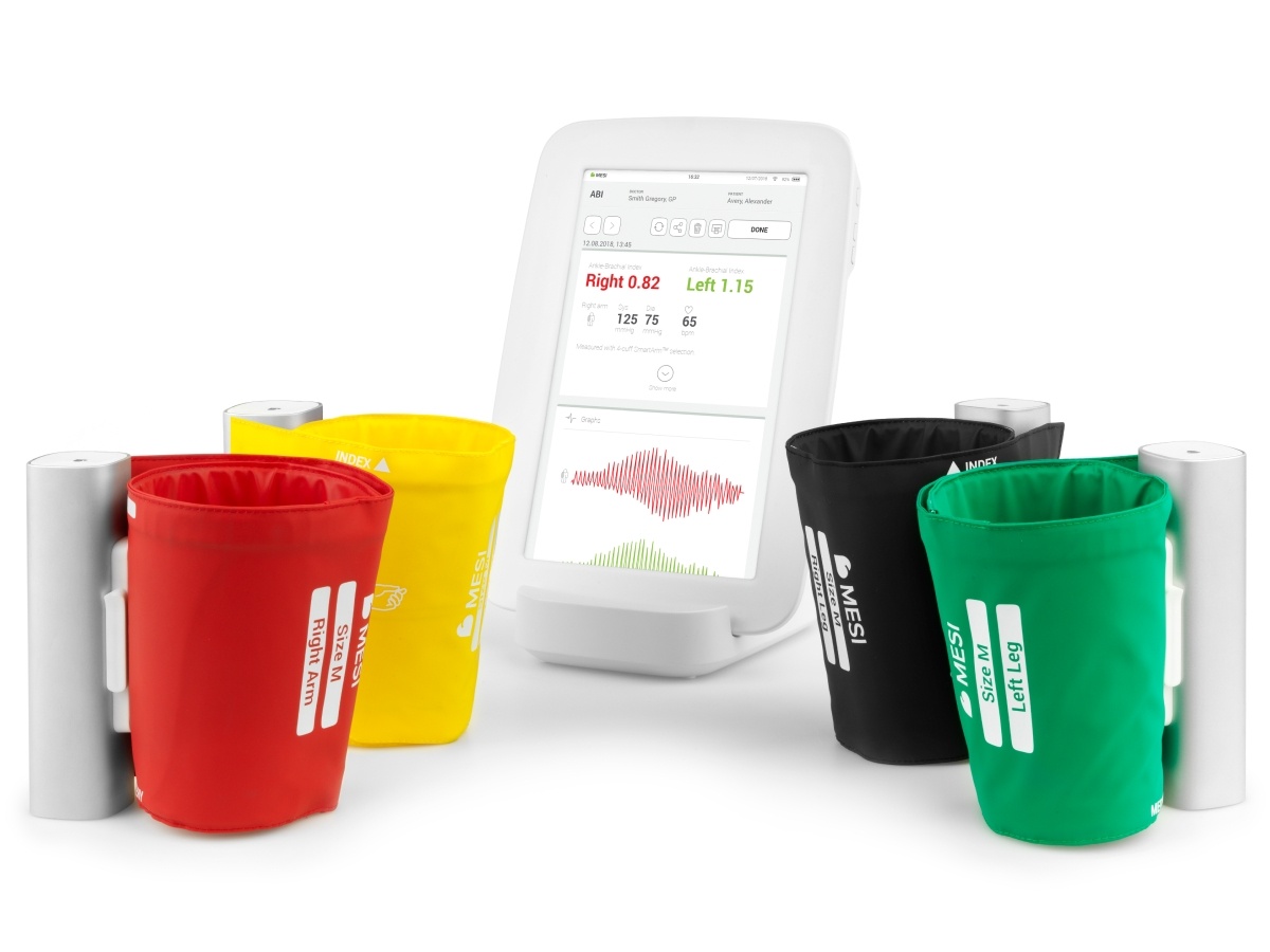
Cuff inflation: Inflatable cuffs are placed on the limbs and inflated to temporarily occlude arterial blood flow.
Oscillation detection: As the cuff slowly deflates, pressure sensors detect small oscillations in cuff pressure caused by arterial pulsations beneath the cuff.
Waveform recording: These oscillations are recorded as pulse volume waveforms (PVR), which reflect changes in arterial blood volume within the limb segment during each cardiac cycle.
Pressure determination: Algorithms analyze the oscillation pattern to determine systolic pressure and calculate the ankle-brachial index (ABI).
Because the cuff captures pulsations from all arteries within the limb segment, the recorded signal reflects the combined perfusion of the arterial network, including the posterior tibial and dorsalis pedis arteries at the ankle. [4][6][7].
Why cuff-based systems cannot display the difference between PT and DP
A fundamental principle of physics and vascular anatomy makes vessel-specific measurement physiologically impossible with any circumferential cuff-based system:
Circumferential pressure transmission: When an ankle cuff inflates, air pressure is transmitted uniformly around the entire ankle circumference, compressing all soft tissues and vessels within that segment. [4][12]
Composite signal detection: During deflation, the pressure sensor detects oscillations from whichever artery produces the strongest or earliest pulse signal — typically the artery with the highest systolic pressure or largest caliber. [4][6]
No spatial resolution: Unlike a handheld Doppler probe that can be physically positioned over individual vessels, a cuff has no mechanism to selectively interrogate one artery while excluding another at the same anatomical level. [4][12]
Anatomical reality: The PT and DP arteries lie at different positions around the ankle (medial vs. anterior/dorsal), but both are compressed simultaneously by the cuff; the resulting oscillometric signal represents the dominant perfusion, not a separable measurement of each vessel. [6][12]
Any vendor claiming to provide separate PT and DP values from a single ankle cuff measurement without independent verification is making a claim inconsistent with the fundamental physics of oscillometry[4].
Alignment with Medical Guidelines and CPT 93922
Guideline-Concordant ABI Calculation
The 2024 AHA/ACC Guidelines on the Management of Lower Extremity Peripheral Artery Disease explicitly endorse automated oscillometric ABI measurement as a Class 1 recommendation for PAD detection[8][13]. The standard protocol for ABI calculation, whether using Doppler or automated methods, is:[4][6][8]
ABI=Higher of (PT or DP systolic pressure at ankle) / Higher of (left or right brachial systolic pressure)
Guidelines do not require reporting separate ABI values for PT and DP; rather, they instruct clinicians to use the higher ankle pressure from either artery, as this represents the best available perfusion to the foot[4][6][8]. This approach:
Accounts for anatomical variation (some patients have dominant PT circulation, others dominant DP)
Improves detection of clinically significant PAD by ensuring the most representative ankle pressure is used
Reflects physiologic foot perfusion, which can be maintained through either major arterial pathway. [8][13]
Why "At Distal Posterior Tibial and Anterior Tibial/Dorsalis Pedis Arteries" means ankle-level Measurement
The CPT 93922 descriptor language - "ankle/brachial indices at distal posterior tibial and anterior tibial/dorsalis pedis arteries" - should be interpreted as defining the anatomical measurement level, not mandating vessel-selective resolution[1][2][3]. Here's why:
Anatomical location specification: The phrase identifies where the measurement is performed (the distal leg/ankle, where both PT and DP are present), not a requirement that each artery be isolated independently[1][3]
Volume plethysmography explicitly listed: CPT 93922 names volume plethysmography as one of three acceptable modalities. If the code required true vessel-by-vessel separation, it would by definition exclude the very modality it explicitly permits — an internally inconsistent interpretation[1][2][3]
Consistency with guideline methodology: AHA/ACC guidelines define ABI measurement at "the ankle" using "the higher of PT or DP" pressures, treating the ankle as a single measurement level with two potential arterial sources[8][13]
Regulatory precedent: FDA clearances for automated ABI plethysmographs (including MESI mTABLET, K201046) use identical language ("measurement of ankle-brachial pressure index... at dorsalis pedis and posterior tibial artery") and are approved specifically for oscillometric methods that inherently measure composite ankle pressure[7][11]
The most physiologically and clinically coherent reading is that "at PT and DP arteries" describes the ankle measurement zone, and a cuff placed at this level capturing the dominant perfusion signal fulfills the code requirement - particularly when paired with the required physiologic waveform (PVR)[1][2][3][9].
How MESI mTABLET Delivers 93922 Compliance
MESI mTABLET ABI performs ABI measurement in two steps: an initial bilateral arm measurement to determine the reference brachial pressure, followed by a simultaneous 3-cuff measurement on the selected arm and both ankles for ABI calculation [7]:
| 93922 Element[1][2] | MESI mTABLET Output[4][6][7] |
|---|---|
| Bilateral brachial pressures | Highest arm pressure used as ABI reference; SmartArm™ identifies higher arm[4][6][7] |
| Ankle pressures at PT/DP level | Conical ankle cuffs at PT/DP territory; algorithm detects highest systolic pressure[4][6][7] |
| ABI calculation | Left and right ABI: highest ankle / highest brachial per leg [4][6][7] |
| Volume plethysmography | PVR at ankle during cuff deflation. [4][5][7][8] |
| TBI (when indicated) | TBI for incompressible arteries (diabetes, calcification)[5][6] |
FDA validation: MESI mTABLET ABI (K201046) cleared as substantially equivalent (21 CFR 870.2780, Class II plethysmograph) for rapid ABI + pulse volume plethysmography in PAD screening, wound care, and compression therapy evaluation[7][11].
Clinical Evidence: Automated ABI/TBI vs Doppler
Peer‑reviewed studies validate MESI's accuracy against Doppler ABI:
| Study | Population | Key Results |
|---|---|---|
| Span et al. (2016)[4] | 136 GP patients | R=0.61; mean diff 0.06±0.14; sens 85%/spec 96% (cut‑off 1.0); 7× faster |
| Watson et al. (2022)[5] | 1860 patients | Sens 57–74%/spec 85–99% (ABI≤0.9); 89–94%/92–96% at 1.0 |
| Fendrik et al. (2023)[6] | 117 patients | r=0.672; optimal 0.99; ABI+TBI detected 92.4% PAD limbs vs imaging |
Takeaway: Automated ABI + PVR (per 2024 AHA/ACC PAD Guideline COR 1) shows small bias vs Doppler and ≥85–90% sensitivity/≥90–96% specificity at optimized cut‑offs[4][5][6][8][13].
Reimbursement guidelines for MESI mTABLET in the USA
Medicare coverage: No NCD; governed by MAC LCDs/Articles for noninvasive vascular studies[9][10].
• Medical necessity: Symptoms (claudication, rest pain, ulcers) or risk factors; ABI + physiologic testing (PVR/waveforms) required[9][10][8]
• Documentation: Numerical ABI, PVR waveforms, interpretation; MESI auto‑generates the report with the option to add comments and printo[4][7]
• Personnel: Varies by MAC (eg RVT supervision)[9]
Commercial payers: Align with CMS LCDs[10]. Modifiers: -52 (unilateral), -26 (professional), -TC (technical)[9].
Liability protection: MESI produces 93922‑level physiologic study (ABI + PVR), equivalent to Doppler per evidence/FDA; aligns with AHA/ACC guidelines[8][13][14].
Bottom line for clinicians: MESI mTABLET enables accurate PAD screening supporting 93922 billing - bill confidently with the reports it generates.