Pulse Wave Velocity (PWV): The hidden marker of arterial age
When we think about cardiovascular risk, we often focus on cholesterol, blood pressure readings, or BMI. But there’s another measurement that has quietly become one of the most powerful predictors of cardiovascular morbidity and mortality: Pulse Wave Velocity (PWV).
PWV doesn’t just tell you what your blood pressure is right now, it also tells you how old your arteries really are.
What is Pulse Wave Velocity or PWV?
Every time the heart beats, it sends a pressure wave through the arterial walls. PWV measures how fast that wave travels between two points in the arterial tree, expressed in meters per second (m/s).
Here’s the key clinical insight: the stiffer the artery, the faster the wave travels. Healthy, elastic arteries absorb and dampen the pressure wave, slowing it down. Stiff, aged arteries, often a result of arteriosclerosis, transmit the wave much faster.
This makes PWV a direct, physical measurement of arterial stiffness, and by extension, a window into vascular age.
Why does PWV matter clinically?
PWV has been extensively studied as an independent predictor of cardiovascular events, separate from traditional risk factors. Large meta-analyse [1] found that each 1 m/s increase in carotid-femoral PWV is associated with a 12–14% higher risk of cardiovascular mortality and events, regardless of other risk factors a patient may have.
Importantly, this predictive value is strongest in low-risk and younger individuals, exactly the patients where standard risk scores tend to underestimate true cardiovascular risk. A large individual-participant meta-analysis [2] found that the hazard ratio for cardiovascular events was highest in people under 50 years old and progressively decreased with age.
This means PWV can flag elevated risk in patients who might otherwise look “normal” on standard checks - patients whose arteries are aging faster than their chronological age would suggest, often before any symptoms appear.
For clinicians, this translates into three practical benefits:
early detection of arterial wall damage, before it becomes symptomatic;
a direct marker of arterial stiffness, reflecting the true elasticity (or lack of it) in the vessel walls and
an “arterial age” estimate, giving a tangible way to communicate vascular health to patients and to motivate lifestyle change.
How is PWV measured?
There are two main types of PWV used in clinical practice, and it’s worth understanding the difference.
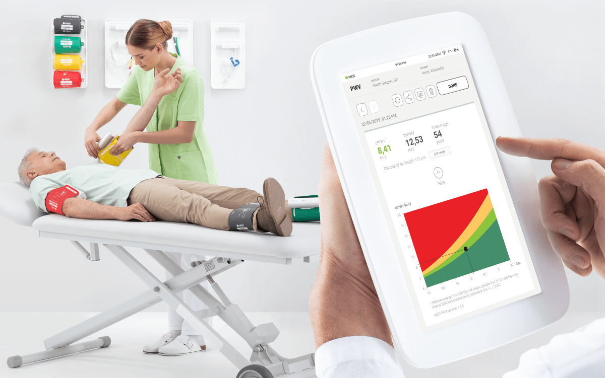
Carotid-Femoral PWV (cfPWV)
cfPWV measures the pulse wave as it travels between the carotid artery (neck) and the femoral artery (inguinal area). This pathway covers the aorta, the body’s largest and most elastic artery, and the one most affected by age-related stiffening.
cfPWV is widely regarded as the reference standard for assessing aortic stiffness and is the measurement most extensively validated in large population studies and referenced in cardiovascular guidelines.
Brachial-Ankle PWV (baPWV)
baPWV measures the pulse wave between the brachial artery (upper arm) and the ankle, covering a longer path that includes both central (aortic) and peripheral arterial segments.
It is technically simpler to perform and is also recognised as a relevant measurement of arterial stiffness. baPWV can be measured using cuffs placed on the arms and ankles, similar to a blood pressure measurement, without the need for direct vessel palpation or specialist training. This makes it considerably easier to implement in routine clinical workflows, particularly in primary care and nursing settings, while still correlating well with cardiovascular risk. A large individual-participant meta-analysis found that for each 1 m/s increase in baPWV, the risk of cardiovascular events increases by 12%, cardiovascular mortality by 13%, and all-cause mortality by 6% [1].
In short: cfPWV is often considered the gold standard for research and central aortic stiffness, while baPWV offers a more practical, scalable option for everyday clinical screening, especially when combined with other vascular assessments.
What do the guidelines say?
Both the 2023 ESH Guidelines for the management of arterial hypertension [3] and the 2024 ESC Guidelines for the management of elevated blood pressure and hypertension [4] recognize arterial stiffness, measured by cfPWV or baPWV, as a marker of hypertension-mediated organ damage (HMOD).
According to the 2023 ESH Guidelines, HMOD assessment is particularly important in individuals with high-normal blood pressure or grade 1 hypertension, especially those who appear to be at low risk, because the result can influence the decision of whether, and how quickly, to start blood-pressure-lowering treatment. HMOD assessment is recommended both at diagnosis and during follow-up, to help evaluate the effectiveness of therapy.
The 2024 ESC Guidelines go a step further, providing concrete diagnostic thresholds for arterial-related HMOD is > 10 m/s for cfPWV and > 14 m/s for baPWV.
The guidelines specify that this kind of optional HMOD testing is most valuable for:
Reclassifying individuals with elevated blood pressure from low-to-moderate to sufficiently high cardiovascular risk to warrant consideration of BP-lowering treatment
Identifying patients who would benefit most from lower BP treatment targets - helping overcome therapeutic inertia in borderline cases (BP or risk near thresholds, masked or white-coat hypertension, non-traditional risk factors)
Risk reclassification in individuals with elevated BP or hypertension under 40 years old
To put this in perspective: out of 1,000 patients seen in a typical primary care setting, roughly 300 will have hypertension and another 150 will sit in the elevated BP range (SBP 130–139 or DBP 80–89 mmHg). For the first group, a high PWV reading can help overcome therapeutic inertia and prompt treatment initiation. For the second group, patients who might otherwise not be treated, a high PWV result can be the deciding factor for starting hypertension therapy.
PWV as part of routine arterial assessment
PWV shouldn’t be viewed in isolation. It fits naturally alongside other vascular assessments, such as Ankle-Brachial Index (ABI) and Toe-Brachial Index (TBI), as part of a broader picture of a patient’s arterial health. PWV is also being explored beyond hypertension management, with emerging evidence linking arterial stiffness to conditions such as pre-eclampsia and to predicting response to interventions like renal denervation.
Together, these measurements help clinicians:
Detect peripheral arterial disease (PAD) and arterial stiffness early
Track vascular changes over time in patients with hypertension, diabetes, or chronic kidney disease
Support more informed decisions in chronic disease management and preventive care
Making PWV part of everyday practice
Historically, PWV measurement required specialized equipment and additional training. This created a barrier for many primary care and nurse-led settings. Automated, integrated devices are changing that, allowing PWV to be captured as part of a routine arterial assessment, alongside other measurements, with standardized protocols and automatically generated reports for trend tracking over time.
The MESI mTABLET PWV makes arterial age measurement fast, automated, and effortlessly integrated into the same wireless, EHR-connected workflow you already use for ABI and other vascular assessments, with no specialist training needed.
Learn more about PWV with MESI mTABLETArterial stiffness develops silently, but it doesn’t have to go undetected. Adding PWV to your routine vascular assessment can help identify at-risk patients earlier and support more proactive, personalized care.